In the diagnosis of lower back pain, the following steps are involved:
1. General history: In this step of diagnosis, the information like the time of beginning of the pain, the reason behind the occurrence of the pain, the agents which can make pain better and worse, etc. are collected.
2. Physical examination: In this method the range of movement of lower back is examined by the use of the movements like straight leg raising and cross leg rising. By doing so the portions getting sensation by these movements are observed and hence the presence of lower back pain is estimated. When there is weakness or numbness in the lower part of the body i.e. the legs it may be due to the lower back problem because during the problem one or the other nerve is compressed and the particular muscle which is compressed leads to problem in the particular area of the leg to which it roots. This is known as the neurological inspection of the lower limbs. In the physical examination the persistence of pain in the regions of hip and sacro-iliac joints are also observed.
3. Blood test: in this type of diagnosis, different blood tests are done to determine different components of blood. The tests include ESR, full blood components count, determination of Rheumatoid factor for Rheumatoid arthritis, etc. blood is also cultured for the recognition of vertebral osteomyelitis.
4. If the doctor suspects the presence of skeletal tuberculosis, Mantoux test can also be performed.
5. The urine test of the patient is also done for the analysis of different components and it is also cultured to detect the presence of any kind of urinary infections.
6. Radiological investigations: In this type of diagnosis the lower back part is subjected to different types of radiological tests like:
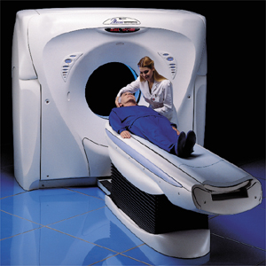
a) CT scan- it uses X- rays to produce 3D images of the scanned area in the form of 5mm thick slices and when these slices are put together one can see the image together to some extent. When the CT scan is done for the spine they give the perfect image of the bones but it is hard to figure out the soft tissues like tumors or herniated discs.
b) Lumbar spine films- in this technique simple x-ray imaging of the affected area is done which provides the image of the alignment of the spine in a 2D manner. Any slippage or fracture in the spine can easily be observed. Tumor eating up the bone can also be seen by this technique as many bone defects are visible.
c) MRI scan: in this technique magnetism is used rather than X-rays for the imaging purpose. The slices obtained in this scan are more serial than the images of CT scan. The can also be taken any other planes like sagittal and coronal than the axil ones. In addition to these features the MRI also gives the details of soft tissues which were not there in the technique of CT scan.
Many other radiological tests like Bone density tests, Discogram, EMG/NCV studies, post myelographic CT scans, myelograms, etc. are also done for the diagnosis of the lower back pain depending upon the condition of the patient.

No comments:
Post a Comment